Table of Contents
Optical Sensing Instruments
Optical Coherence Tomography (OCT)
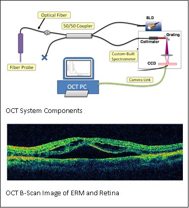
OCT provides very high resolution (micron scale) images of anatomical structures within the tissue. Within Ophthalmology, OCT systems typically perform imaging through microscope optics to provide 2D cross-sectional images (“B-mode”) of the retina. These systems are predominantly used for diagnosis, treatment planning, and in a few cases, for optical biopsy and image guided laser surgery. We are developing intra-ocular instruments that combine surgical function with OCT imaging capability in a very small form factor.
 Dr. Kang’s group has built a custom common path Fourier domain Optical Coherence Tomography (CP-OCT) designed specifically for interfacing with smart surgical instruments for vitreoretinal surgery. The system can be used for retinal tissue imaging and also as a depth range sensor. The CP-OCT works on the principle that coherent light interferes with itself after it is reflected and refracted. This interference encodes (in Fourier domain) the distances from the reference plane to the optically opaque layers of the imaged sample. The whole axial depth scan (A-Scan) can be collected in a single exposure and processed with Fast Fourier Transform.
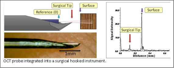
 OCT imaging can be used as a range finder by extracting the distance from the probe’s reference to the first large peak in the A-Scan, which generally represents the surface of the sample. Resulting real-time range information is used as a feedback parameter in a virtual fixture framework for cooperative robot control. In the case of targeting, additional retinal tracking ability is required. This may be provided by video processing to extract tool position relative to the retina.
- Safety Barrier: The system enforces a safety constraint to prevent the probe from approaching the target surface closer than a specified threshold distance. The robot moves freely within the workspace to comply with forces exerted by the user on the control handle, with the exception of the forbidden boundary sensed via the OCT. For example, this “virtual wall” is reached when the tip of the instrument is located ~150μm from the sample surface.
ML45iQB2oU0
- Surface Tracking: This gives the operator the ability to scan the probe across a surface while maintaining a constant distance offset. One intention for the surface tracking is to compensate for retinal motion due to respiratory function.
FufTkqaws4c
- Targeting: This is a complex behavior that assists the surgeon to place the pick over a subsurface target identified in a scan and then penetrate the surface to hit the target in a semiautonomous fashion.
Status
We have built a robust OCT system and associated micro instruments. The device is compatible with our EyeSAW software framework allowing for rapid prototyping of “behaviors” combining the functionality of other EyeSAW devices. We have added audio sensory substitution to indicate the proximity of the instruments to the retina. Another method uses video overlays to present OCT images.
Future
We are looking into building new OCT integrated instruments and more intelligent ways to present the data to the surgeons. Another research area is OCT image processing to detect structures such as blood vessels and membranes that can be used for robot assisted interventions.
Project Personnel
| JHU Whiting School | JHU Hospital, Wilmer Eye Institute |
|---|---|
| Dr. Russell Taylor | Dr. James Handa |
| Dr. Greg Hager | Dr. Peter Gehlbach |
| Dr. Jin Kang | |
| Dr. Iulian Iordachita | |
| Marcin Balicki | |
| Xuan Liu | |
| Yi Yang |
Funding
- NIH - 1 R01 EB 007969-01 A1
- NSF - EEC9731748
- JHU - Internal funds
Affiliated labs
Publications
- Xuan Liu, Marcin Balicki, Russell H. Taylor, and Jin U. Kang. “Automatic online spectral calibration of Fourier-domain OCT for robot-assisted vitreoretinal surgery” , in SPIE Advanced Biomedical and Clinical Diagnostic Systems IX,25 January 2011.
- Xuan Liu, Marcin Balicki, Russell H. Taylor, and Jin U. Kang. “Towards Automatic Online Calibration of Fourier-Domain OCT for Robot-Assisted Vitreoretinal Surgery” Optics Express Journal 2010. Also appeared in Virtual Journal for Biomedical Optics V.6, I.1, 1/2011.
- “Single Fiber Optical Coherence Tomography Microsurgical Instruments for Computer and Robot-Assisted Retinal Surgery” Marcin Balicki, Jae-Ho Han, Iulian Iordachita, Peter Gehlbach, James Handa, Jin Kang, Russell Taylor. Proceedings of the MICCAI Conference (Oral Presentation, 5% acceptance rate), March 2009. Best Paper Award in Computer Assisted Intervention Systems and Medical Robotics (September 2009)
- Jae-Ho Han, Marcin Balicki, Kang Zhang, Jae-Ho Han, Marcin Balicki, Kang Zhang, Xuan Liu, James Handa, Russell Taylor, and Jin U. Kang. “Common-path Fourier-domain Optical Coherence Tomography with a Fiber Optic Probe Integrated Into a Surgical Needle” Proceedings of CLEO Conference, May 2009