Table of Contents
Intraoperative Image-Guided TransOral Robotic Surgery
TransOral robotic surgery (TORS) is a minimally invasive surgical intervention to treat oropharyngeal cancer, including resection of base of tongue tumors. It is a progressively more viable option in addressing a national trend, where cancers associated with the human papilloma virus (HPV) has resulted in younger patients (40 to 60 years), whose treatment options must equally consider cure and longer term quality of life. TORS offers an increasingly attractive treatment modality with minimal morbidity and reduced toxicity compared to conventional regimens of chemo+radiotherapy. Despite encouraging oncologic and functional results, remaining barriers include a lack of useful landmarks, especially with infiltrative, submucosal base of tongue cancers, as well as distorted operative patient position (mouth open + tongue extended) not reflected in pre-operative images. The goal of this project is the development of image-guidance with stereoscopic video augmentation on the da Vinci S System for base of tongue tumor resection in transOral robotic surgery (TORS). We propose using cone-beam computed tomography (CBCT) to capture intraoperative patient deformation from which critical structures (e.g., the tumor and adjacent arteries), segmented from preoperative CT/MR, are deformably registered to the intraoperative scene. Augmentation of TORS endoscopic video with image and planning data defining the target and critical structures offers the potential to improve navigation, spatial orientation, confidence, and tissue resection, especially in the hands of less experienced surgeons.
Methods
Video Augmentation: Stereoscopic video augmentation is achieved by extending the cisst/ SURGICAL ASSISTANCE WORKSTATION (SAW), open source libraries developed at the Engineering Research Center for Computer Integrated Surgery at the Johns Hopkins University, to support a modular architectural design.
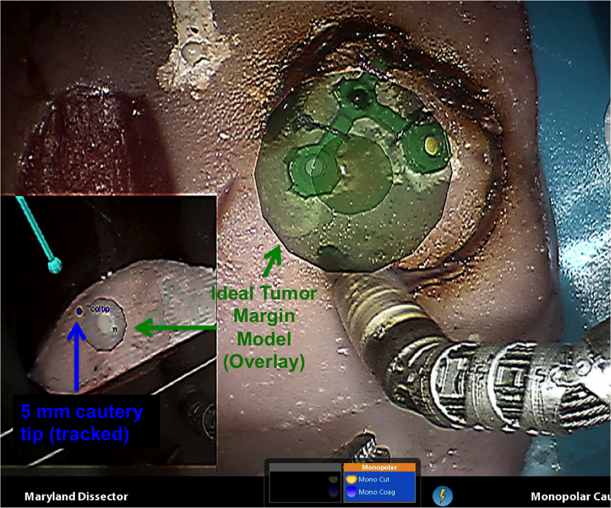
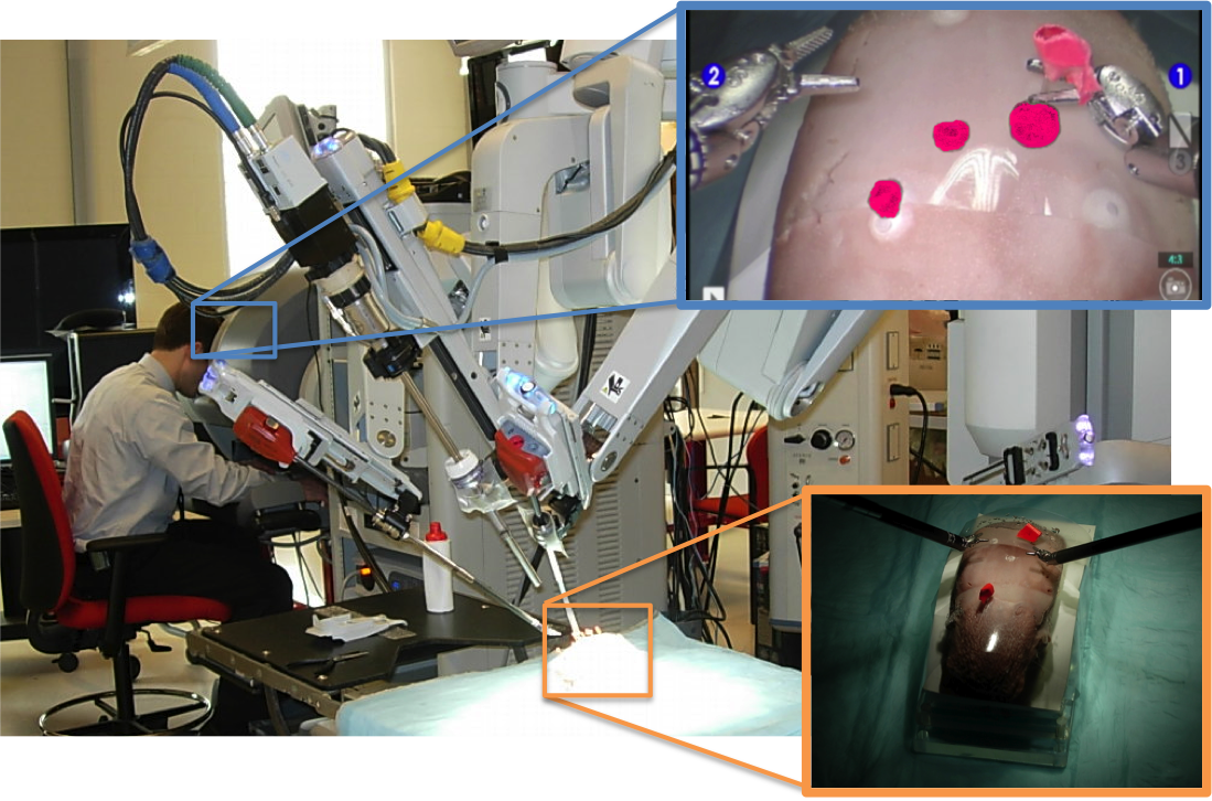 Figure 1. Photograph capturing image guidance system as seen during a dry lab experiment at Hackerman Hall, Mock OR.
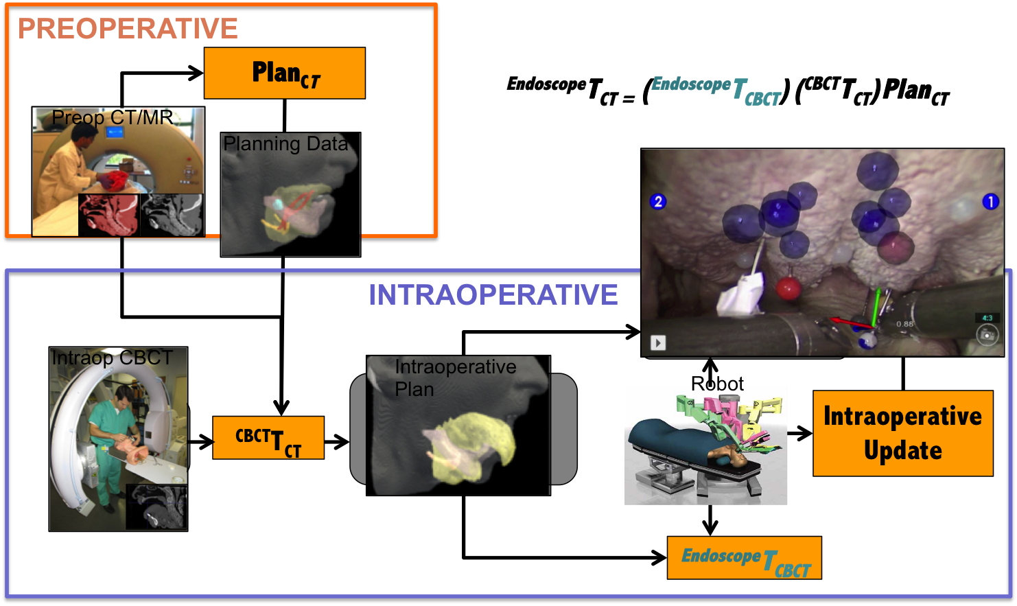
Intraoperative Workflow: Intraoperative CBCT registers critical information derived from standard preoperative diagnostic CT/MRI to integrated image guidance through augmented reality.
Figure 2. Diagram of proposed image guidance workflow
Status and Results to Date
Phantoms: Using ex-vivo porcine tongues, cadaveric and in-vivo pig phantoms we have show statistically significant advantages using intraoperative image data and variations of image-guidance/video augmentation on target localization in TORS.
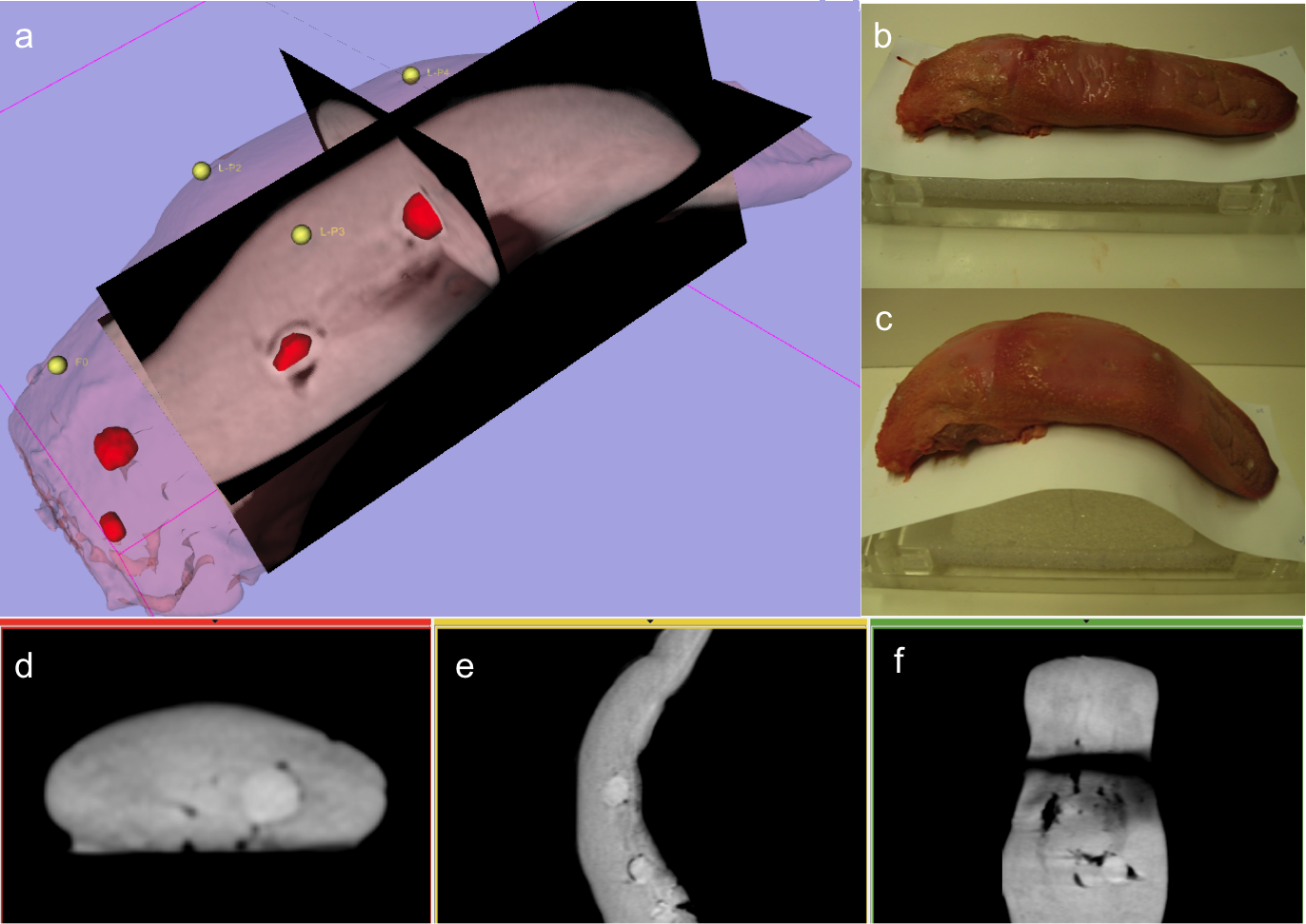
Figure 4. Excised dry tongue phantom
Figure 5. Invivo pig phantom
Results:
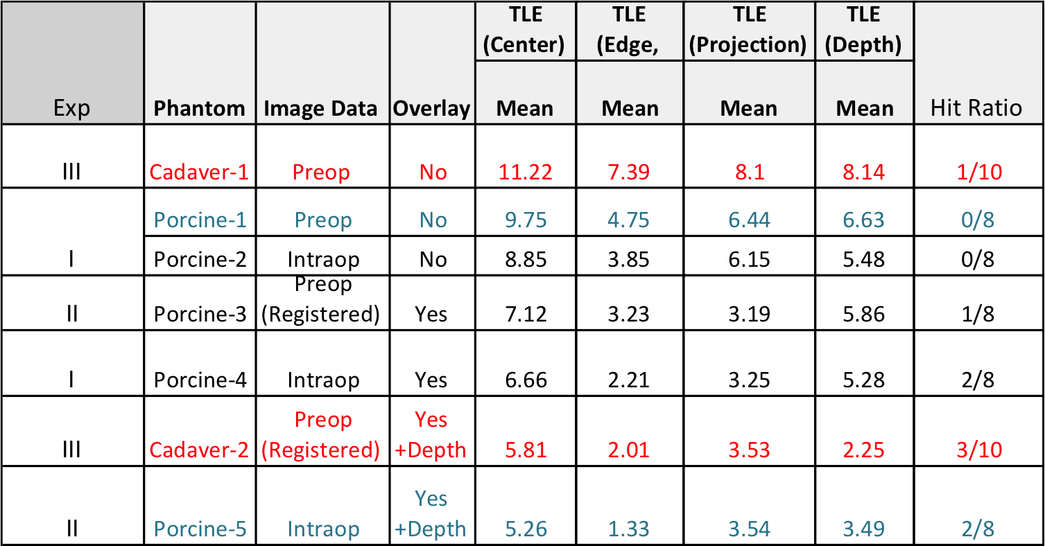
Table 1. Results show improved TLE (center) between simulated current workflow and proposed image-guidance with statistical significance comparing porcine-1 to porcine-5 (p-value = 0.0151) & cadaveric-1 to cadaveric-2 phantoms (p-value = 0.0189) [in submission]
Funding
- Intuitive Surgical Inc.
- NIH
- Swirnow Family Foundation
Affiliated labs
Project Personnel
| Name | Institution | Affiliation |
|---|---|---|
| Dr. Russell Taylor | Johns Hopkins University | Principal Investigator |
| Dr. Jeffrey Siewerdsen | Johns Hopkins University | Principal Investigator |
| Dr. Jeremy Richmon | Johns Hopkins University | Clinician |
| Dr. Jonathon Sorger | Intuitive Surgical Inc. | Research Scientist |
| Sureerat Reaungamornrat | Johns Hopkins University | Graduate student |
| Wen P. Liu | Johns Hopkins University | Graduate student |
Publications
- W. P. Liu, S. Reaugamornrat, A. Deguet, J. M. Sorger, J. H. Siewerdsen, J. Richmon, R. H. Taylor, “Toward Intraoperative Image-Guided Transoral Robotic Surgery”, Journal of Robotic Surgery, July 2013.
- Reaungamornrat S, Liu WP, Wang AS, Otake Y, Nithiananthan S, Uneri A, Schafer S, Tryggestad E, Richmon J, Sorger JM, Siewerdsen JH, and Taylor RH, “Deformable registration for cone-beam CT-guided transoral robotic base of tongue surgery,” Phys. Med. Biol. 2013.
- J. Richmon, W. P. Liu, R. H. Taylor, S. Reaugamornrat,J. Sorger, J. H. Siewerdsen, “Intraoperative Image-Guided Transoral Robotic Surgery: Pre-Clinical Studies”,20th International Federation of Oto-Rhino-Laryngological Societies (IFOS) 2013
- S. Reaugamornrat, W. P. Liu, S. Schafer, Y. Otake, S, Nithiananthan, A. Uneri, J. Richmon, J. Sorger, J. H. Siewerdsen, R. H. Taylor, “Deformable Registration for Cone-Beam CT Guidance of Robot-Assisted, Trans-Oral Base-of-Tongue Surgery” in Society of Photographic Instrumentation Engineers (SPIE) Medical Imaging Conference, 2013."Deformable Registration for Cone-Beam CT Guidance of Robot-Assisted, Trans-Oral Base-of-Tongue Surgery"
- W. P. Liu, S. Reaugamornrat, A. Deguet, J. M. Sorger, J. H. Siewerdsen, J. Richmon, R. H. Taylor, "Toward Intraoperative Image-Guided TransOral Robotic Surgery" [Hamlyn Symposium, London UK 2012]
