Tabiscope: Mobile Device Camera Connector

Last updated: 2014/05/09 23:42

Summary

In resource poor third world nations there is a lack of affordable technology for visualization and picture-taking during endoscopy. The standard endoscope imaging tower is extremely expensive and may not be easily purchased. A commercial solution is available for first world countries with the Endoscope-I; however, it is limited to using recent generations of iPhones which have limited screen size that reduces the clinical usefulness of this visualization during endoscopy. In addition, the high cost of video towers and iPhones is a major barrier for wide-spread use, especially outside of first-world nations or in resource poor communities. Thus, our goal is to create an Android-based application and adapter to be compatible with endoscopes in order to address these existing problems.

  • Students: Daniel Ahn (dahn9@jhu.edu), Deepak Lingam (dlingam1@jhu.edu), Kyle Wong (wong.kyle5@gmail.com)
  • Mentor(s): Kevin Olds (kolds1@jhu.edu), Dr. Amit Kochhar (akochha4@jhmi.edu), Dr. Simon Best(sbest2@jhmi.edu)

(Photo Courtesy of http://endoscope-i.com/)

Background, Specific Aims, and Significance

The lack of a low cost solution for capturing digital images during endoscopy leads to painful, costly, and uncomfortable re-examinations. This is creates a barrier to sharing clinical information regarding the endoscopy, and required additional time for clinicians to see and examine the patients themselves, some of whom may be in emergency situations. In rural communities, this is worsened since specialists such as Otolaryngologists, are not always available. Thus, after a first examination, the patient must return when a physician is available or travel to a neighboring city. The lack of stored digital images also prevents clinicians from easily tracking a patient’s health progress over time.

Additional applications for this technology would be for other surgical specialties that use endoscopes such as urology, obstetrics and gynaecology, gastroenterology, and orthopedic surgery. Having a universal adapter would permit this. Furthermore, having this tablet adapter would enable clinicians to take pictures during endoscopy on the hospital floor and in the Emergency Room even if they do not have iPhones or the standard endoscope imaging tower.

Old Deliverables

  • Minimum: (3/30)
    1. A working adapter for a specific Android tablet for a rigid endoscope
    2. Android application with a Graphical User Interface (GUI) for adjusting tablet's camera settings and saving pictures to the device
  • Expected: (4/15)
    1. A working adapter for flexible endoscopes as well (to have universal adapter for all endoscopes)
    2. Android application with GUI for organizing images by patient identifier
  • Maximum: (5/2)
    1. Universal adapter for connecting any tablet to any endoscope
    2. Portable light source that ensures high quality images
    3. Android application that uploads and offers secure viewing of patient endoscopy images

Updated Deliverables

  • Minimum: (4/6)
    1. Adapter for a Specific Tablet for Endoscope (Done)
    2. Android app: Being able to view images using the tablet’s camera (Done)
  • Expected: (4/21)
    1. Updated Adapter that allows users to adjust tablet position (Done)
    2. Android app: GUI / label images (patient identifier) (In Progress)
    3. Automatic detection of image circle and maximization of ROI (In Progress)
  • Maximum: (5/2)
    1. Android application that uploads and offers secure viewing of patient endoscopy images
    2. Real-time image processing methods to prevent specular reflection
    3. Portable light source that ensures high quality images
    4. Universal adapter for connecting any camera to any endoscope

Technical Approach

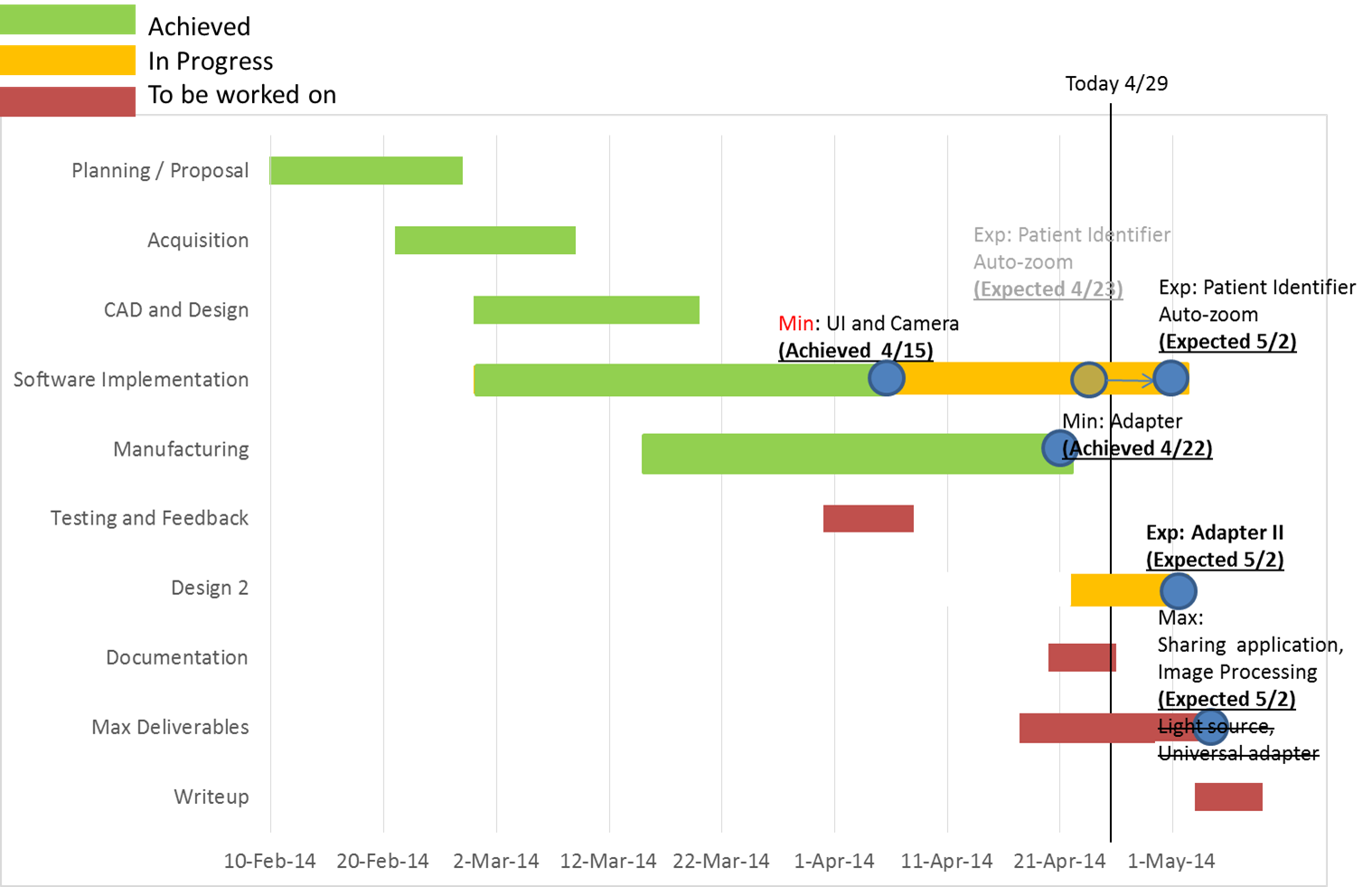
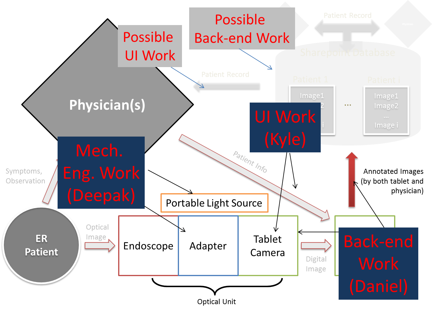
In order to address this problem, we hope to create an application for Android devices using their open source technology for taking high quality pictures and saving, organizing, and transferring the pictures easily and securely. We also seek to create an adapter to attach an external camera directly to the endoscope and stream the video real-time to an Android tablet. It may be necessary to create an optical component of the adapter in order to obtain images of usable quality. We may also need to have a portable light source for using the tablet’s camera with the endoscope. Once we have a working adapter for a single camera, the goal would be to create a more universal adapter to fit any other camera.

GUI Class Diagram: guiclassdiagram.pdf

Standard Endoscope Eyepiece size: 32 mm

Dependencies

  • Android tablet with a high-resolution camera (DONE)
    1. plan A: borrow and use one from the Johns Hopkins Outpatient Center Head and Neck Surgery Department - follow-up with Dr. Kochhar
    2. plan B: receive money to buy an Android tablet - follow-up on Dr. Kochhar and Dr. Best
    3. plan C: use personal Android phone for initial testing
  • A functional endoscope (DONE)
    1. plan A: borrow or get an old endoscope from the Johns Hopkins Outpatient Center Head and Neck Surgery Department
    2. follow-up with Dr. Kochhar
    3. plan B: borrow the old endoscope that Kevin Olds currently has
  • Access to a machine shop or 3D printer for manufacturing an adapter
    1. plan A: have a highly detailed 3D CAD model of the adapter to be built using 3D printer in the DMC
    2. plan B: get access to any of the JHU Mechanical Engineering Machine Shop
    3. plan C: ask the machinist in the WSE Machine Shop to manufacture our design
    4. plan D: ask friends who have access to machine shops to manufacture our design
  • Access to mentors (DONE)
    1. schedule weekly meetings with Kevin Olds: Tuesday 2:45pm @Robotorium
    2. send out email updates every two weeks
    3. schedule monthly meetings with Dr. Kochhar and Dr. Best
    4. get optics / lens advice from Dr. Kang (and other contacts through Kevin)

Milestones and Status

  • Minimum Deliverable 1: Adapter for a Specific Tablet for Endoscope
    • Planned Date: 3/30/2014
    • Expected Date: 4/06/2014
    • Status: Achieved (4/22/2014)

<WRAP center round box 60%> </WRAP>

  • Minimum Deliverable 2: Android app: Being able to view images using the tablet’s camera
    • Planned Date: 4/15/2014
    • Expected Date: 4/21/2014
    • Status: Achieved (4/15/2014)

<WRAP center round box 60%> </WRAP>

  • Expected Deliverable 1: Adjustable tablet adapter
    • Planned Date: 4/21/2014
    • Expected Date: 5/2/2014
    • Status: Achieved (5/2/2014)

<WRAP center round box 60%> </WRAP>

  • Expected Deliverable 2: Android app: GUI / label images (patient identifier)
    • Planned Date: 4/23/2014
    • Expected Date: 5/2/2014
    • Status: In Progress
  • Expected Deliverable 3: Android app: Automatic detection of image circle and maximization of ROI
    • Planned Date: 4/23/2014
    • Expected Date: 5/2/2014
    • Status: In Progress

<WRAP center round box 60%> </WRAP>

  • Milestone name: Maximum Deliverables
    • Planned Date: 5/02/2014
    • Status: Awaiting Start

Reports and presentations

Project Bibliography

  • Portable Light Source
    • Sznitman, Raphael, et al. “Active multispectral illumination and image fusion for retinal microsurgery.” Information Processing in Computer-Assisted Interventions. Springer Berlin Heidelberg, 2010. 12-22.
  • Related Patents
    • Matsumoto, Seiji, Etsuo Nakano, and Suwao Sato. “Battery-powered light source arrangement for endoscope.” U.S. Patent No. 6,260,994. 17 Jul. 2001.
    • Irion, Klaus. “Endoscope with LED illumination.”
    • Shipp, John I. “LED illumination system for endoscopic cameras.” U.S. Patent No. 6,449,006. 10 Sep. 2002.
  • Circle Detection
    • Duda, Richard O., and Peter E. Hart. “Use of the Hough transformation to detect lines and curves in pictures.” Communications of the ACM 15.1 (1972): 11-15.
  • Specular Reflection Removal
    • Nayar, Shree K., Xi-Sheng Fang, and Terrance Boult. “Separation of reflection components using color and polarization.” International Journal of Computer Vision 21.3 (1997): 163-186.
    • Oh, JungHwan, et al. “Informative frame classification for endoscopy video.” Medical Image Analysis 11.2 (2007): 110-127.
    • Stehle, Thomas. “Removal of specular reflections in endoscopic images.” Acta Polytechnica 46.4 (2006).
    • Kaup, André, Katrin Meisinger, and Til Aach. “Frequency selective signal extrapolation with applications to error concealment in image communication.” AEU-International Journal of Electronics and Communications 59.3 (2005)

Other Resources and Project Files

Source Code

GitHub Repository
Contact Daniel Ahn or Kyle Wong for access.

CAD Files

Unfortunately the wiki does not allow CAD file extensions to get uploaded.
Contact Deepak Lingam for files.

Lab Safety Certificates

Paper Seminars

Deepak Lingam

Kyle Wong

Daniel Ahn

Meeting Notes

courses/446/2014/446-2014-07/mobile_device_camera_connector.txt · Last modified: 2019/08/07 16:01 by 127.0.0.1


ERC CISST    LCSR    WSE    JHU