Contact Us
CiiS Lab
Johns Hopkins University
112 Hackerman Hall
3400 N. Charles Street
Baltimore, MD 21218
Directions
Lab Director
Russell Taylor
127 Hackerman Hall
rht@jhu.edu

Please allow the contents of this page to remain confidential until our technical paper is published (expected by 07/01/2014)
Last updated: 05/09/2014 Latest: Summary Report Uploaded
A capacitor model of the knee joint is developed to measure joint width in CT volumes. The implementation takes the form of a gpu-enabled MATLAB library.


Certain anatomical variations in knee morphology (e.g. tibial slope, etc) has recently been found to correlate with increased risk of injury resulting from load-bearing activities. This is significant for combat infantry personnel deployed abroad, who routinely carries a pack of up to 100 lbs while on active duty. In order to attempt to establish a valuable predictor of such risks, an efficient and accurate way of characterizing knee morphology from high quality CT data is needed. Joint space narrowing is also indicative of progression of diseases such as osteoarthritis, which affects nearly 27 million people in the US alone. The development of a robust method to detect such changes in joint space morphology is the primary aim of this project.
Goals:

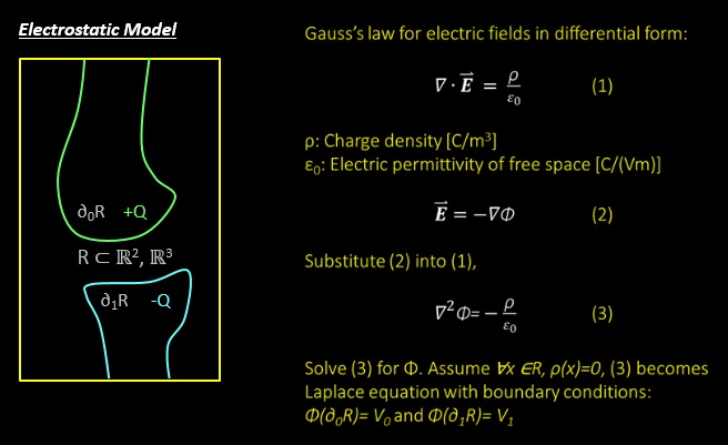
The laws of physics gives an elegant solution to the problem in the form of Maxwell's equations. The femoral and tibial surfaces can be modeled as two surfaces of a capacitor each applied with a positive and negative voltage, respectively.
All dependencies are met.

* here list references and reading material
[1] J. R. Giffin, T. M. Vogrin, T. Zantop, S. Woo, and C. D. Harner, “Effects of Increasing Tibial Slope on the Biomechanics of the Knee,” Am. J. Sports Med. 32(2): 376-382 (2004).
[2] J. Hashemi, N. Chandrashekar, B. Gill, B. D. Beynnon, J. R. Slauterbeck, R. C. Schutt Jr., H. Mansouri, and E. Dabezies, “The Geometry of the Tibial Plateau and Its Influence on the Biomechanics of the Tibiofemoral Joint,” J. Bone and Joint Surg. 90: 2724-2734 (2008).
[3] J. Hashemi, N. Chandrashekar, H. Mansouri, B. Gill, J. R. Slauterbeck, R. C. Schutt Jr., E. Dabezies, and B. D. Beynnon, “Shallow Medial Tibial Plateau and Steep Medial and Lateral Tibial Slopes : New Risk Factors for Anterior Cruciate Ligament Injuries,” Am. J. Sports. Med. 38: 54-52 (2010).
[4] S. G. McLean, S. M. Lucey, S. Rohrer, and C. Brandon, “Knee joint anatomy predicts high-risk in vivo dynamic landing knee biomechanics,”Clin. Biomech. (2010) (in press doi:10.1016/j.clinbiomech.2010.06.002).
[5] K. B. Shelburne,H.-J. Kim, W. I. Sterett, and M. G. Pandy, “Effect of Posterior Tibial Slope on Knee Biomechanics during Functional Activity,” Orth. Res. Soc. Feb. 223-231 (2011).
[6] S. J. Shultz and R. J. Schmitz, “Tibial Plateau Geometry Influences Lower Extremity Biomechanics During Landing,” Am. J. Sports Med. 40(9): 2029-2036 (2012).
[7] W. Zbijewski, P. De Jean, P. Prakash, Y. Ding, J. W. Stayman, N. Packard, R. Senn, D. Yang, J. Yorkston, A. Machado, J. A. Carrino, and J. H. Siewerdsen, “A dedicated cone-beam CT system for musculoskeletal extremities imaging: Design, optimization, and initial performance characterization,” Med. Phys. 38(8): 4700 - 4713 (2011).
[8] A. Muhit, S. Arora, M. Ogawa, W. Zbijewski, J. W. Stayman, G. Thawait, N. Packard, R. Senn, D. Yang, J. Yorkston, C. Bingham, K. Means, J. A. Carrino, and J. H. Siewerdsen, “Peripheral Quantitative CT (pQCT) Using a Dedicated Extremity Cone-Beam CT Scanner,” SPIE Proc. Medical Imaging 2013, Orlando FL.
[9] Yezzi, A. J., & Prince, J. L. (2003). An Eulerian PDE approach for computing tissue thickness. IEEE Transactions on Medical Imaging, 22(10), 1332–9. doi:10.1109/TMI.2003.817775
Source files are stored on lab servers and are backed up daily. They are available upon request (qcao@jhmi.edu).