Contact Us
CiiS Lab
Johns Hopkins University
112 Hackerman Hall
3400 N. Charles Street
Baltimore, MD 21218
Directions
Lab Director
Russell Taylor
127 Hackerman Hall
rht@jhu.edu

Last updated: May 5, 2016
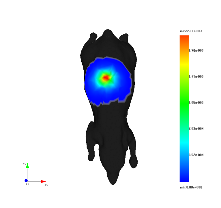
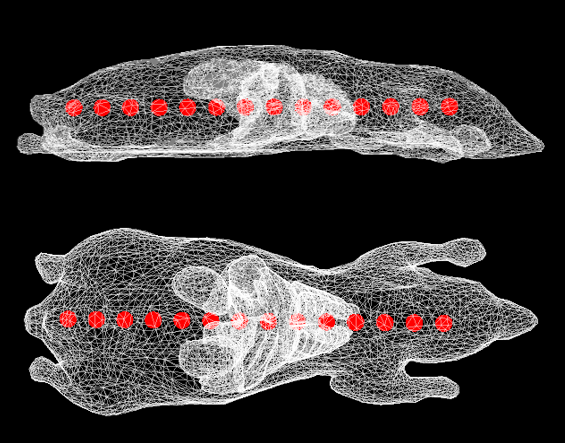
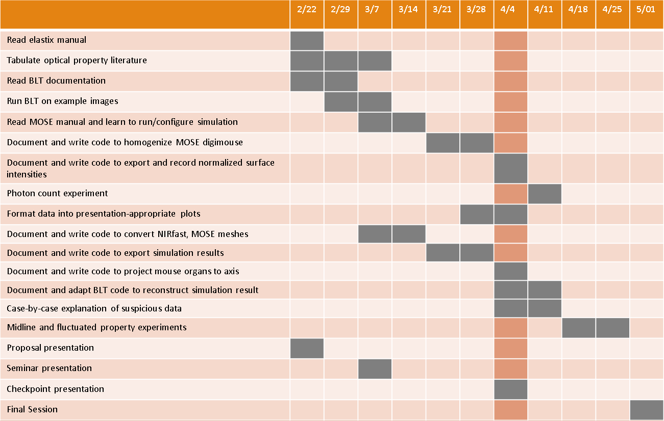
Project 15 is to gather literature values for the optical properties of mouse organs for incorporation into bioluminescence tomography (BLT) reconstruction. Furthermore, this project aims to investigate the effects of mouse heterogeneity on the accuracy of center-of-mass (COM) reconstruction by using the Molecular Optical Simulation Environment (MOSE) to solve the forward light transport problem for a heterogeneous mouse. Project 15 also aims to produce an automatic mouse segmentation process and to perform experiments with physical light source implants in mice. Update: the automated mouse segmentation and physical implant experiments are no longer planned.
The Small Animal Radiation Research Platform (SARRP) is a preclinical research tool for mouse CBCT imaging and radiation delivery. In order to improve radiation targeting of low-contrast tissues, BLT functionality has been implemented for the SARRP. Previous experiments involved physical light source implants in relatively homogenous region of the mouse (abdomen), and the reconstruction was performed under assumption that the body homogeneously bears the properties of adipose tissue. Nonetheless, center of mass (COM) targeting accuracy of 1 mm has been achieved using the homogeneity assumption. Information about the optical properties of mouse organs and their spatial distribution of those organs may help improve BLT reconstruction results. Project 15 aims to augment the existing BLT workflow through the following aims:
Update: the automated segmentation and physical implant experiments are no longer planned.
Status: all dependencies have been met.
Project Background
Elastix
Molecular Optical Simulation Environment (MOSE)
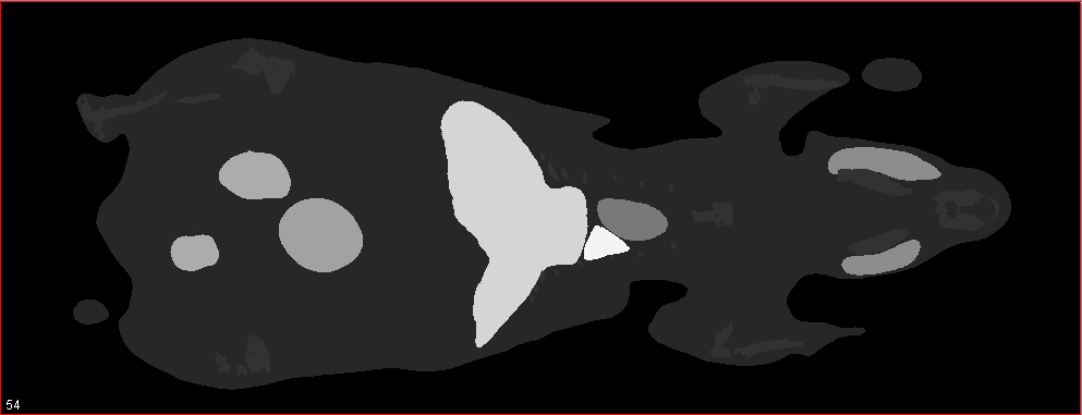
Image Segmentation
Optical Properties
Here give list of other project files (e.g., source code) associated with the project.