Table of Contents
X-ray Image Based Navigation for Hip Osteotomy
Last updated: 03-01-2012, 3:30PM
Summary
- Students: Jesse Hamilton, Mike Van Maele
- Mentor(s): Mehran Armand, Yoshito Otake, Ryan Murphy
Periacetabular osteotomy (PAO) is a joint reconstruction surgery for increasing femoral head coverage and improving stability in patients with developmental dysplasia of the hip (DDH). Our research mentors have previously developed a software package for geometrical and biomechanical planning of PAO called the Biomechanical Guidance System (BGS). One of its key features is intraoperative fragment tracking using a Polaris optical system. However, because of the wider availability of C-arm imagers in hospitals and surgeon familiarity with fluoroscopy, an implementation of BGS utilizing x-ray navigation is desired.
The first aim of this project is to develop a protocol and software pipeline for an X-ray image guided navigation system for performing PAO. The second objective is to compare the proposed pipeline with the current procedure, which utilizes optical tracker navigation. The proposed procedure involves placing several metallic radiopaque BBs on (1) the uncut pelvis to provide a virtual reference frame, and (2) the bone fragment undergoing realignment. Prior to surgery, two C-arm images will be acquired at different poses, de-warped, and registered to a preoperative CT model. A small (3x3x5 cm) fluoroscopic tracker (FTRAC) will be attached to the pelvis to recover image pose. Following fragment realignment, two more C-arm images will be acquired and registered to the CT model. The BGS software will display biomechanical and radiographic data to help the surgeon evaluate whether the realignment has met the preoperative plan.
Background, Specific Aims, and Significance
DDH is characterized by a malformed acetabulum, or hip socket, exhibiting insufficient coverage of the femoral head and high joint contact pressure. Such elevated pressure can lead to complications such as degenerative osteoarthritis and fracture of the acetabular labrum [Millis 1995]. DDH is most prevalent in young women, often under the age of 30. Hip arthroplasty is not recommended in patients who are young and active or have unilateral dysplasia. These patients are likely to outlive mechanical joint replacements and require revision surgery, and there are concerns regarding prosthetic surface durability and preservation of bone stock. For these individuals, reconstructive surgeries are preferable to prosthetics.
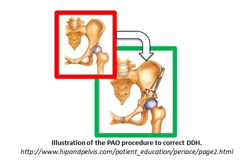
One effective joint reconstruction surgery is periacetabular osteotomy (PAO), which reorients the acetabulum to increase femoral head coverage and lower joint contact pressure. In particular, the Ganz osteotomy [Ganz 1988] utilizes a single incision and four osteotomies, and it preserves the patient’s posterior column and vascular supply.
Our research mentors have previously developed a software package for computer-assisted PAO named the Biomechanical Guidance System (BGS) which features both preoperative planning and intraoperative fragment tracking [Murphy 2010]. The BGS procedure begins with acquisition of a full pelvic CT scan and manual segmentation of the femur and pelvis. The Lunate-Trace algorithm is used to segment and generate a surface mesh model of the acetabulum, and radiographic angles are measured from the CT image. During the planning stage, the software computes contact pressures via Discrete Element Analysis, and it suggests a reorientation of the acetabulum that minimizes simultaneous peak contact pressure in sitting, standing, and walking positions [Murphy 2010].
The current BGS system is capable of intraoperative fragment tracking using a Polaris 3D position sensor. After performing a pivot calibration, a dynamic rigid body (DRB) is attached to the pelvis to serve as a fixed reference frame. A gross model-to-patient registration is performed by touching a surgical tool tip (also equipped with a DRB) to four known anatomical locations on the pelvis, and an iterative closest point algorithm performs fine surface registration. The surgeon drills four confidence points on the ilium to serve as a fixed reference frame. After the osteotomy, the surgeon updates the model-to-patient registration by touching the four confidence points on the ilium and the four landmark points on the realigned fragment. BGS displays radiographic angles and biomechanical data indicating how the fragment moved during surgery. If necessary, the surgeon can reorient the fragment and update the registration until the desired outcome is achieved.
As mentioned in the Summary, an X-ray based navigation system for the BGS-assisted PAO would likely appeal to surgeons by relying on the imaging modality with which they are accustomed to working. Moreover, the availability of optical trackers in a typical OR is not likely to be as widespread as the availability of a C-arm imager.
Deliverables
- Minimum: (Expectedby 03-30-2012)
- (COMPLETED) Develop a surgical pipeline/protocol from acquiring pre-op CT to displaying post-op biomechanics.
- (COMPLETED) Decide on a method for firmly attaching BBs to bone.
- (COMPLETED) Develop code to manually classify BBs in images as confidence, fragment, or FTRAC
- (COMPLETED) Compare of experimental results obtained from mock surgery using (i) proposed method (x-ray navigation) and (ii) conventional method (optical tracker navigation).
- Expected: (Expected by 04-27-2012)
- (COMPLETED) Investigate non-rigid attachment of FTRAC
- (NOT COMPLETED) Integrate x-ray navigation procedure with existing BGS software.
- (COMPLETED) Compare of experimental results obtained from cadaveric surgery using (i) proposed method (x-ray navigation) and (ii) conventional method (optical tracker navigation).
- Maximum: (Expected by 05-04-2012)
- (IN PROGRESS) Develop a robust technique for automatically initializing the ECM algorithm
- (NOT COMPLETED) Investigate alternatives to FTRAC.
- (NOT COMPLETED) Simulate error analysis of in-image fiducial.
Technical Approach
Procedure Outline
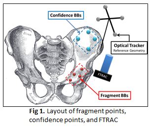
We propose a novel protocol and software pipeline for an x-ray guided navigation system for performing PAO. First the surgeon acquires a preoperative CT scan of the patient’s hip region. Next BGS constructs a 3D biomechanical model of the hip and femur and assists in planning the osteotomy. Intraoperative fragment tracking will be achieved using metallic, radiopaque BBs. It should be noted that this is similar to an FDA approved practice for radiostereometry analysis, in which tantalum beads are injected onto bone for tracking in certain orthopedic applications [RSA Biomedical 2009]. In our procedure, four BBs are placed (1) on the operative side of the pelvis to serve as a fixed reference frame (called the “confidence points”) and (2) on the acetabular fragment (called the “fragment points”. The BBs should be placed 3 to 4 centimeters apart and not be coplanar.
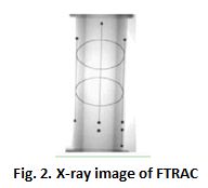
The surgeon also attaches a fluoroscopic tracker (FTRAC) [Jain 2005] to the pelvis for estimating the image pose. The FTRAC is a fiducial containing points, lines, and ellipses in a configuration that allows the 6DOF C-arm pose to be uniquely calculated from any view (Fig. 2). The FTRAC is non-rigidly placed within the field of view of the C-arm before acquiring images. As long as the FTRAC remains stationary with respect to the patient while acquiring a set of pre-operative or intra-operative images, it is possible to recover C-arm pose.

The raw fluoroscopic images are warped due to the curved shape of the detector and interference from the Earth’s magnetic field. In addition, this distortion varies with C-arm position. We utilize existing software to correct this distortion. A metallic grid phantom is placed on the detector and a single image is acquired preoperatively. A distortion map is created by fitting a fifth degree Bernstein polynomial to the data and interpolating over all pixels in the image. Future work may investigate principal component analysis (PCA) based correction, which has the added advantage of yielding pose-dependent distortion maps [Chintalapani 2007].
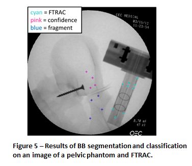
After rigid fixation of the BBs, non-rigid attachment of the FTRAC, and creation of the distortion maps, two or more C-arm fluoroscopy images are acquired at roughly 30° separation. BBs are segmented using a radial symmetry algorithm and classified as FTRAC, confidence, or fragment points (Fig 3). In the cadaver experiment, larger diameter BBs were used for confidence points compared to fragment points, which permitted size discrimination.
Next the C-arm pose is estimated using the FTRAC. Two pose estimation algorithms were investigated: expectation conditional maximization (ECM) and pose from orthography with scaling (POSIT). POSIT requires prior knowledge of matching feature points between an image and a 3D model, whereas ECM is correspondenceless. However, ECM requires an initial pose estimate, unlike POSIT. The correspondenceless property of ECM makes it an attractive choice for this project.
After pose estimation the FTRAC, confidence, and fragment BBs are backprojected from the x-ray images into a 3-dimensional patient coordinate frame centered on the FTRAC. The transformation from this “patient frame” to the pre-operative CT volume is obtained via 2D-3D registration (for which code was provided by our mentors).
After the osteotomy, the entire imaging workflow is repeated (place FTRAC in field of view, acquire images, classify BBs, estimate C-arm pose, backproject BBs, perform 2D-3D registration). This permits calculation of the transformation which maps pre-realignment fragment points to post-realignment fragment points. Future work should incorporate this information with BGS to update the CT visualization and biomechanical analyses.
Dependencies
- (COMPLETED) Get mock OR access
- (COMPLETED) Schedule meetings with mentors
- (COMPLETED) Get radiation badges
- (NOT NEEDED) Get radiation training
- (COMPLETED) Get access to PCs with 2D-3D registration software and BGS
- (COMPLETED) Get pelvic phantom
Milestones and Status
- Milestone name: Establish Wiki Page
- Planned Date: 2/16
- Expected Date: 2/17
- Status: Begun
- Milestone name: Radiation Training
- Planned Date: 2/24
- Expected Date: 3/05
- Status: Contacted Dr. Granlund and Mina Razavi
- Milestone name: Software Support
- Planned Date: 2/21
- Expected Date: 2/23
- Status: MATLAB Files & Data Sets Acquired
Reports and presentations
- Project Plan
- Project Background Reading
- See Bibliography below for links.
- Project Checkpoint
- Experimental Results
- Paper Seminar Presentations
- Project Final Presentation
- Project Final Report
- links to any appendices or other material
Project Bibliography
- Armand M, Lepisto J, Tallroth K, Elias J, Chao E. Outcome of periacetabular disease. Acta Orthopedica 2005;76(3):303-13.
- Armiger RS, Armand M, Lepisto J, Minhas D, Tallroth K, Mears SC, Waites MD. Evaluation of a computerized measurement technique for joint alignment before and during periacetabular osteotomy. Computer Aided Surgery 2007;12(4):215-24.
- Armiger RS, Armand M, Tallroth K, Lepisto J, Mears SC. Three-dimensional mechanical evaluation of joint contact pressure in 12 periacetabular osteotomy patients with 10-year follow-up. Acta Orthopedica 2009 04;80(2):155-61.
- Chintalapani G, Jain AK, Taylor RH. Statistical characterization of C-arm distortion with application to intra-operative distortion correction. Proc SPIE 2007;6509:65092Y.
- David P, Dementhon D, Duraiswami R, Samet H. SoftPOSIT: Simultaneous pose and correspondence determination. International Journal of Computer Vision 2004 09/15;59(3):259-84.
- Dementhon DF, Davis LS. Model-based object pose in 25 lines of code. International Journal of Computer Vision 1995;15(1):123-41.
- Jain AK, Mustafa T, Zhou Y, Burdette C, Chirikjian GS, Fichtinger G. FTRAC—A robust fluoroscope tracking fiducial. Med Phys 2005 October 2005;32(10):3185-98.
- Kang X, Taylor RH, Armand M, Otake Y, Yau WP, Cheung PYS, Hu Y. Correspondenceless 3D-2D registration based on expectation conditional maximization. Proc SPIE 2011 March 3, 2011;7964(1):79642Z.
- Millis MB, Murphy SB. Osteotomies of the hip in the prevention and treatment of osteoarthritis. The Journal of Bone & Joint Surgery 1995;41:626-47.
- Otake Y, Armand M, Armiger R, Kutzer M, Basafa E, Kazanzides P, Taylor R. Intraoperative image based multi-view 2D/3D registration for image-guided orthopaedic surgery: Incorporation of fiducial-based C-arm tracking and GPU-acceleration. Medical Imaging, IEEE Transactions on 2011.
Other Resources and Project Files
Here give list of other project files (e.g., source code) associated with the project. If these are online give a link to an appropriate external repository or to uploaded media files under this name space.


