Contact Us
CiiS Lab
Johns Hopkins University
112 Hackerman Hall
3400 N. Charles Street
Baltimore, MD 21218
Directions
Lab Director
Russell Taylor
127 Hackerman Hall
rht@jhu.edu
Vendor Independent PA Imaging System Enabled with Asynchronous Laser Source
Last updated: 03/26/2017
Enter a short narrative description here
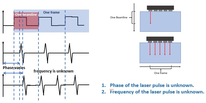
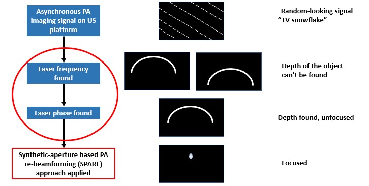
Photoacoustic (PA) imaging is an imaging modality that derives image contrast from the optical absorption coefficient of the tissue being imaged . Owing to its deep penetration, high resolution and safety, it is intensively and widely applied in fundamental, preclinical and clinical studies as a very effective optical imaging modality. However, hardware of PA imaging hinders a universal application of this technology. Implementation either relies on low-efficiency Ultrasound (US) beamformers or vender-variant PA platforms. If conventional US scanner implementing PA imaging is viable, then the cost of PA imaging will be lower so that more life will be saved. Thanks to the effort of Medical UltraSound Imaging and Intervention Collaboration (MUSiiC) Lab, an innovative photoacoustic re-beamforming approach has been developed to make real-time PA implementation on conventional US platforms possible. However, this approach is applied on an ultrasound platform where frame rate and probe’s beamline sweeping rate are manually set. Whereas, conventional clinical ultrasound scanners don’t possess such “triggers” for frames and sweeping. In another word, the phase and frequency of the laser pulse are unknown, leading to a random-looking image where the transmitted signal is asynchronous with the received signal. Hence, a reconstruction method which recovers the laser frequency and the laser phase are significant and crucial for implementing PA imaging on conventional US platform.
To implement PA imaging on US platforms, difference between the two should be well considered and image reconstruction methods for PA imaging are acquired.
 One critical problem that must be solved before PA imaging can be totally applied onto conventional US scanners is the synchronization. Since conventional US platforms don’t have laser trigger, when does each frame starts (phase of the laser) and when does the frequency of the laser are unknown.
 The way to solve the problem is to first simulate the whole process in Matlab, with assistance of k-wave tool box. Then transplant the algorithm in C++ onto Ulterius, construct phantoms and validate on clinical ultrasound platforms.
 One very initial and intuitive though about the algorithm is to set bounds and intervals for Tframe and Tpulse, and search the optimal one by brute-force. Other approaches may include solving optimization problems to get the best image quality.
 Instead of synchronization, image reconstruction has also to be real-time. This has been achieved by group 13 in Computer Integrated Surgery II 2016. The technical approach here is to combine the real-time imaging part with data obtained after synchronization to make PA imaging on US platform viable.
Software:
Hardware:
Here give list of other project files (e.g., source code) associated with the project. If these are online give a link to an appropriate external repository or to uploaded media files under this name space.