Contact Us
CiiS Lab
Johns Hopkins University
112 Hackerman Hall
3400 N. Charles Street
Baltimore, MD 21218
Directions
Lab Director
Russell Taylor
127 Hackerman Hall
rht@jhu.edu

Last updated: May 8th 14:23
Registration of multimodal images is a powerful tool for transferring preoperative plans into an intraoperative environment, thereby enhancing the surgeon’s accuracy to localize instruments in the operating room. Intraoperative ultrasound (US) to preoperative Computer Tomography (CT) registration is often a challenging task due to multiple factors, including the low signal-to-noise ratio of the US images and the presence of artifacts. This work explores Short Lag Spatial Coherence (SLSC) beamforming as an alternative to traditional Delay-And-Sum (DAS) ultrasound images to increase contrast and resolution for US-to-CT registration tasks. . The objective of this work is to explore Short-Lag Spatial Coherence (SLSC) beamforming as an alternative to delay-and-sum (DAS) beamforming to reduce speckle noise and enhance bone boundaries. In addition, we propose Locally Weighted SLSC (LW-SLSC) beamforming to further improve bone segmentation.
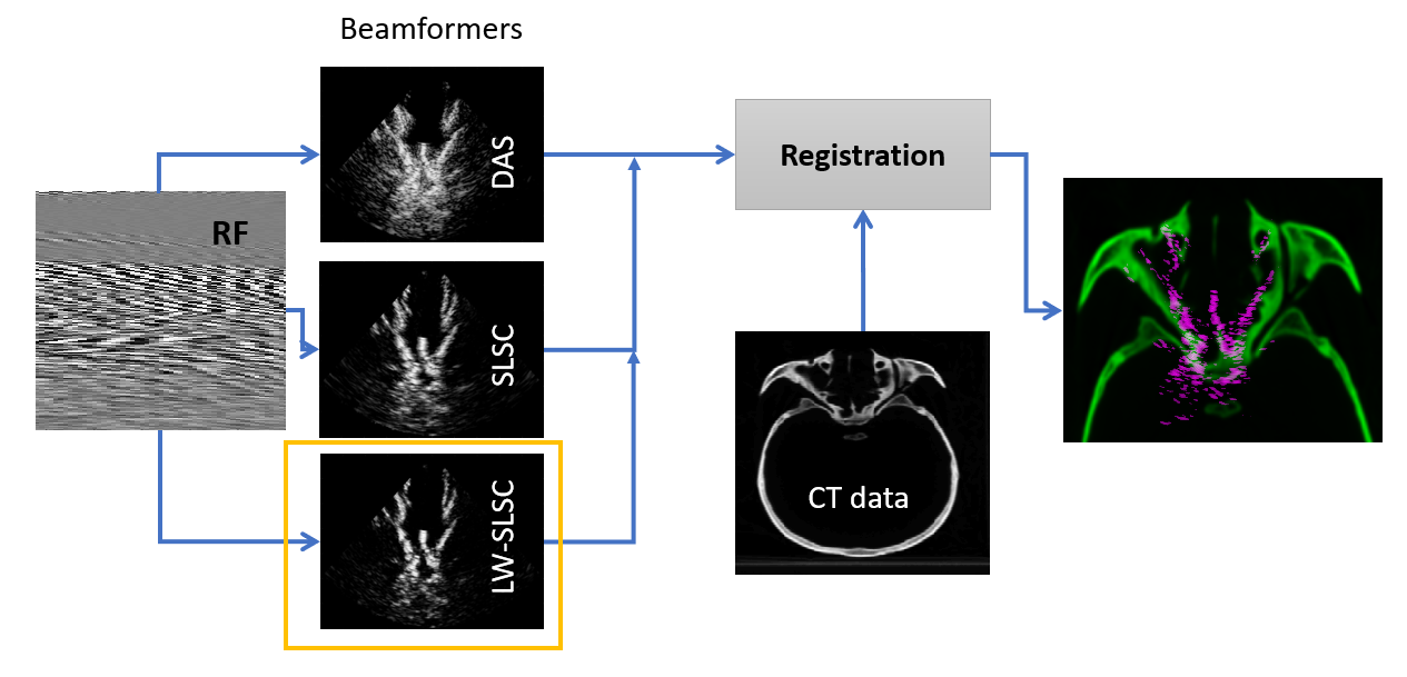
 The overall initial framework is presented in Fig. 1. First, US channel is acquired and processed in offline mode with
several beamforming techniques (DAS, SLSC and LW-SLSC). Then, in order to remove undesired reflection from water
particles and bottom of the tank, segmentation using Fuzzy C-means was conducted. The algorithm divided each
reconstructed US image in 3 regions: bone, water and not-image (due to the US transducer geometry) and selected
the layer of bone only. Then, resampling was performed so the US image feature a similar pixel resolution than the
CT data. Finally, registration was performed using Mattes Mutual information (fixed) as optimizer and least square
method as metric.
The overall initial framework is presented in Fig. 1. First, US channel is acquired and processed in offline mode with
several beamforming techniques (DAS, SLSC and LW-SLSC). Then, in order to remove undesired reflection from water
particles and bottom of the tank, segmentation using Fuzzy C-means was conducted. The algorithm divided each
reconstructed US image in 3 regions: bone, water and not-image (due to the US transducer geometry) and selected
the layer of bone only. Then, resampling was performed so the US image feature a similar pixel resolution than the
CT data. Finally, registration was performed using Mattes Mutual information (fixed) as optimizer and least square
method as metric.
Goal: Explore methods to improve accuracy of US-CT image registration through improved US image resolution
Specific aims:
- Enhance relevant features of US images to improve automatic registration performance: The more well-defined the borders of the desired region in a US image, the better the registration performance.
- Develop a robust US beamformer for such purposes: This will allow to control the quality of the US images in other to better extract relevant features.
- Explore the registration improvement when considering additional information of Photoacoustic (PA) imaging: This will give us insight of the potential of PA imaging to contribute to translation of preoperative to intraoperative information.
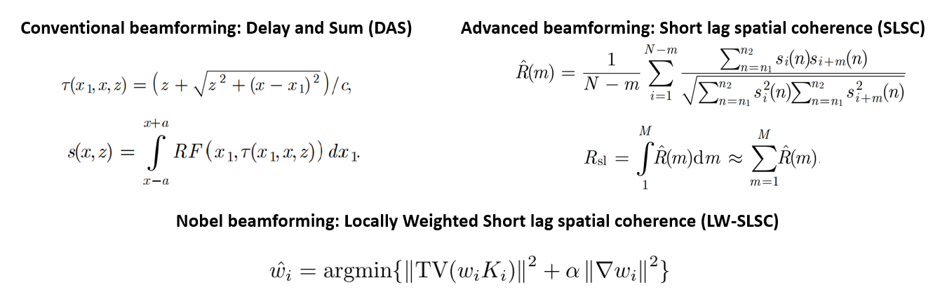
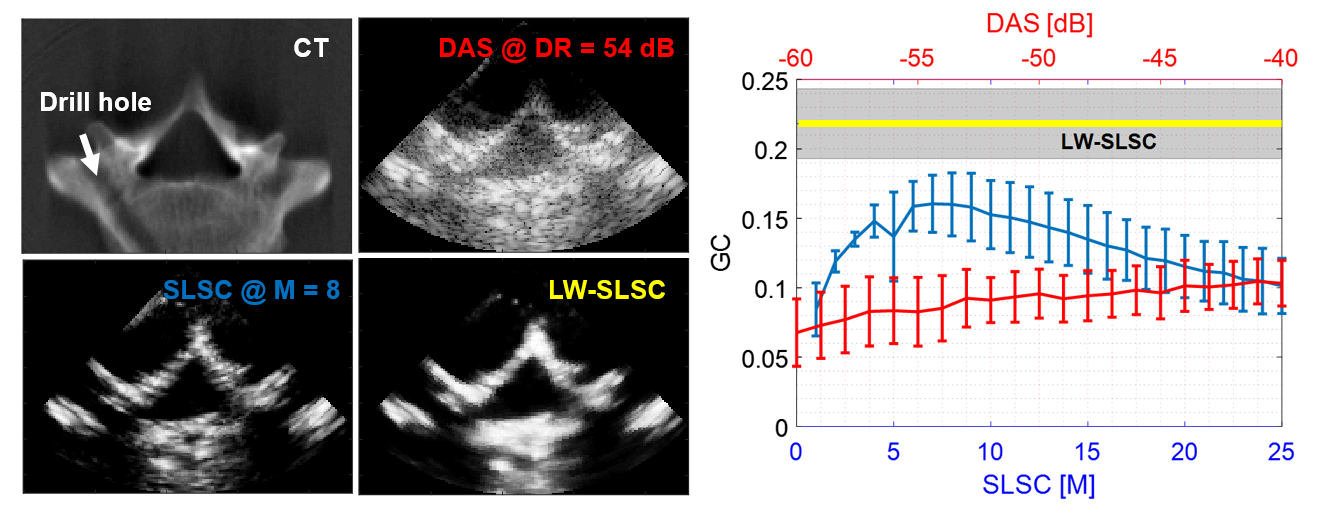
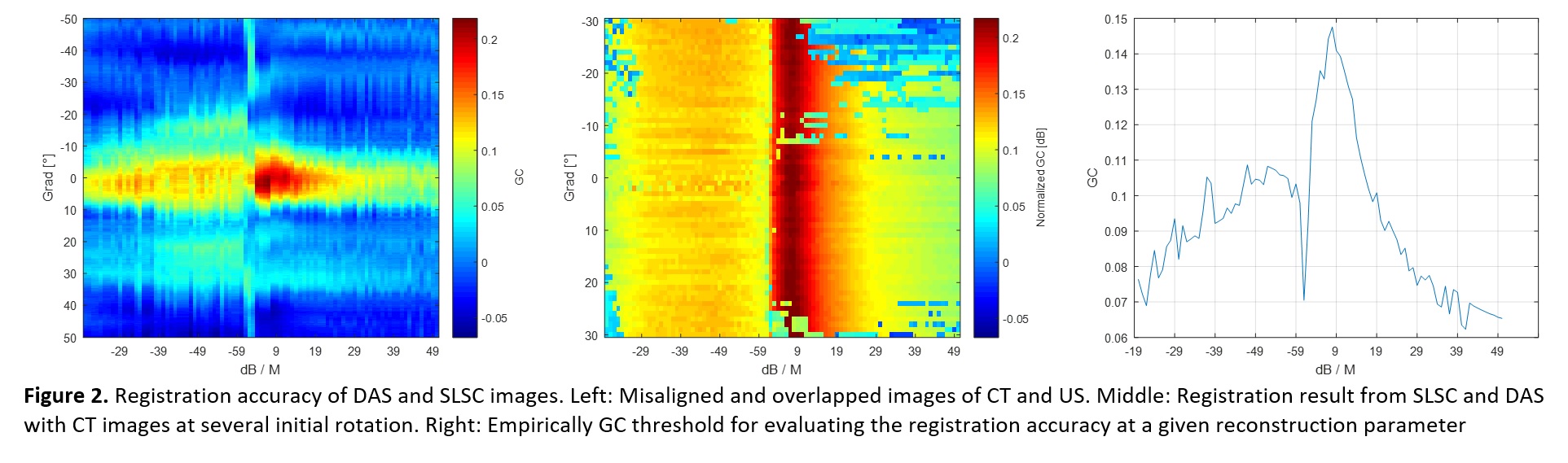
Instead of averaging the cumulative sum up to a lag value M (out of a preselected total of N lags, where M≤N), LW-SLSC beamforming computes the weighted coefficients for N lags by minimizing the total variation of the weighted sum within a moving kernel. In order to preserve the high resolution located at higher lags, this adaptive solution is regularized using the L2-norm with a gradient operator. US channel data was acquired for 10 different views of a human vertebra submerged in deionized water using an Alpinion ECUBE-12R system and SP1-5 phased array probe (3.8 MHz center frequency, 65 mm depth, 50 mm focus). SLSC images were computed with M varying from 1 to 25, and DAS images were created with the dynamic range (DR) varying from -60 to -40 dB. LW-SLSC images were computed with a 1.20 mm (lateral) x 1.92 mm (axial) kernel, N=50, 50% overlap and 0.1 regularized coefficient obtained from L-curve tests. The Gradient Correlation (GC) was measured to evaluate the bone structure similarity of each ultrasound imaging method when compared to CT images.
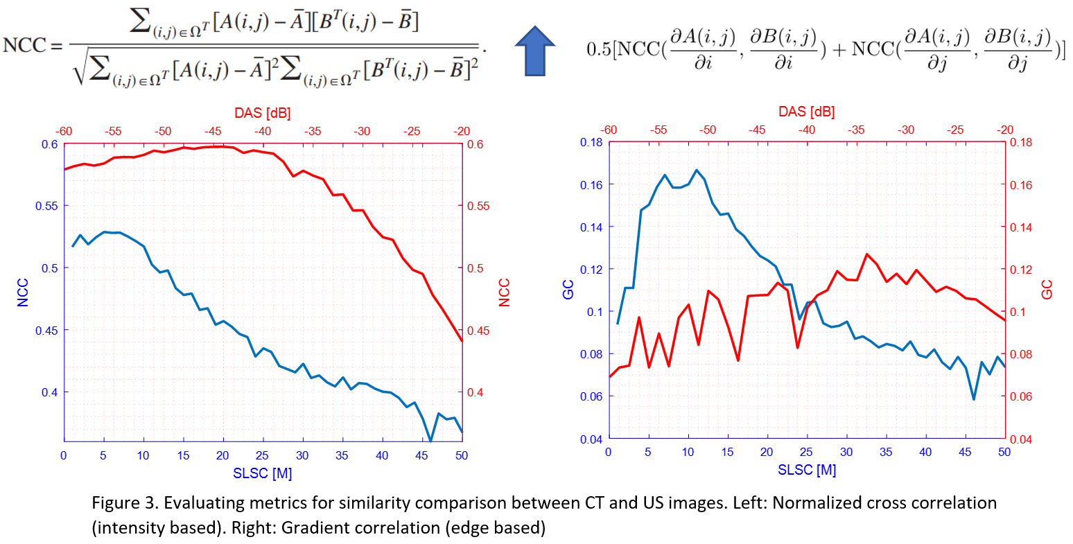
Example CT, DAS, SLSC and LW-SLSC images are shown in Fig. 1 alongside the mean standard deviation of GC measurements. Overall, SLSC outperforms DAS for a range of parameters commonly used in the literature (e.g., M=5-25, DR=-50 to -60 dB) when considering the similarity of bone structures in the CT and US-based images. An additional improvement is observed with LW-SLSC over SLSC (e.g., 8.2 dB mean contrast-to-noise ratio increase, 0.10 mean GC increase)
In order to assess the level of accuracy that the GC factor provides regarding registration and similarity, intensional misalignment (performed manually) of the reconstructed US and CT images was performed from 30 to -30 degrees and then overlapped. Note that no registration step was conducted. From the results, an empirical threshold vs the parameter for DAS and SLSC can be generated around 5 degrees of rotation, which corresponds to GC values less than 0.06. Later, registration of a set of images was conducted when the CT was rotated a certain angle (in the same range of the previous experiment). The empirical threshold previously calculated was in accordance with registration results.
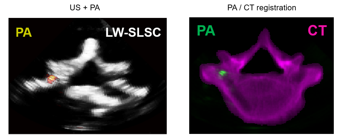
Finally, the same experiment was conducted with the addition of Photo acoustic (PA) signals. We used a caterer operating at 750 wavelength and 70% energy firing to the drill hole cavity inside the vertebra. Then, channel data as for both ultrasound and photo acoustic was acquired. The additional data from PA can be considered as a separate image that is already registered to the US images. Then, applying an advanced beamforming (either SLSC or LW-SLSC) and registering to the CT image, is it possible to track the location of the PA signal. In practice a PA signal can be installed to the tip of an operating tool, which could be tracked inside the human vertebra. However, this is a preliminary result, and more experiments are required for such statement.
Acquisition of CT images of human spine:
- Scheduling use of CT machine in the medical campus with Professor Siewerdsen and/or Homewood campus with Michelle Graham (from PULSE lab)
- Cannot acquire CT myself because did not take the CT training course
- Availability of the spine sample (it should not be an issue)
- Roche et al. “Rigid Registration of 3-D Ultrasound With MR Images: A New Approach Combining Intensity and Gradient Information”, 2001.
- Wein et al. “Simulation and Fully Automatic Multimodal Registration of Medical Ultrasound”, 2007.
- Wein et al. “Automatic CT-ultrasound registration for diagnostic imaging and image-guided intervention”, 2008.
- Bell et al. “Short-Lag Spatial Coherence of Backscattered Echoes: Imaging Characteristics”, 2011.
- Wong et al. “Real-time ultrasound-guided spinal anesthesia using the SonixGPS needle tracking system: a case report”, 2013.
- Shubert et al. “A novel drill design for photoacoustic guided surgeries”, 2017.
Here give list of other project files (e.g., source code) associated with the project. If these are online give a link to an appropriate external repository or to uploaded media files under this name space.2018-20