Contact Us
CiiS Lab
Johns Hopkins University
112 Hackerman Hall
3400 N. Charles Street
Baltimore, MD 21218
Directions
Lab Director
Russell Taylor
127 Hackerman Hall
rht@jhu.edu
The Steady-Hand Eye Robot is a cooperatively-controlled robot assistant designed for retinal microsurgery. Cooperative control allows the surgeon to have full control of the robot, with his hand movements dictating exactly the movements of the robot. The robot can also be a valuable assistant during high-risk procedures, by incorporating virtual fixtures to help protect the patient, and by eliminating physiological tremor in the surgeon's hand during surgery.
The EyeRobot1 (ER1) is a five degree of freedom robot, with three translational axes (x, y, z) and two rotational axes (roll and tilt). As our initial design it is used to develop the understanding of ergonomic and precision requirements for eye surgery.
Virtual RCM (vRCM) is an important cooperative-control element utilized by the ER1 to safely position the surgical tool for retinal microsurgery. The surgeon chooses a point on the eye, usually the insertion point of the surgical tool in the sclera of the eye, to act as the vRCM. The motion of the tool will then be limited only to certain rotations about that one point, helping to protect the patient and prevent possible damage to the eye.
EyeRobotRCM EyeRobotRCM
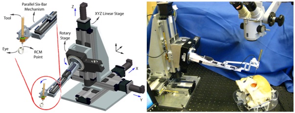
Eye Robot 2 (ER2) is an intermediate design towards a stable and fully capable microsurgery research platform for the evaluation and development of robot-assisted microsurgical procedures and devices.

ER2 manipulator consists of four subassemblies: 1) XYZ linear stages for translation, 2) rotary stage for rolling, 3) a tilting mechanism with a mechanical RCM, and 4) a tool adaptor with a handle force sensor. Parker Daedal 404XR linear stages (Parker Hannifin Corp., Rohnert Park, CA) with precise ball-screw are used to provide 100 mm travel in XYZ axes with a bidirectional repeatability of 3 µm and positioning resolution of 1 µm. A Newport URS 100B rotary stage (Newport Corp., Irvine, CA) is used for rolling, with a resolution of 0.0005° and repeatability of 0.0001°. A THK KR15 linear stage (THK America Inc., Schaumburg, IL) with travel of 100 mm and repeatability of ±3 µm is used to provide tilting motion. The last active joint is a custom-designed RCM mechanism. A 6-DOF ATI Nano17 force/torque sensor (ATI Industrial Au-tomation, Apex, NC) is mounted between the RCM mechanism and the tool handle. The end-effector has a free spinning tool axis for holding surgical instruments.
Here are some improvements over ER1:
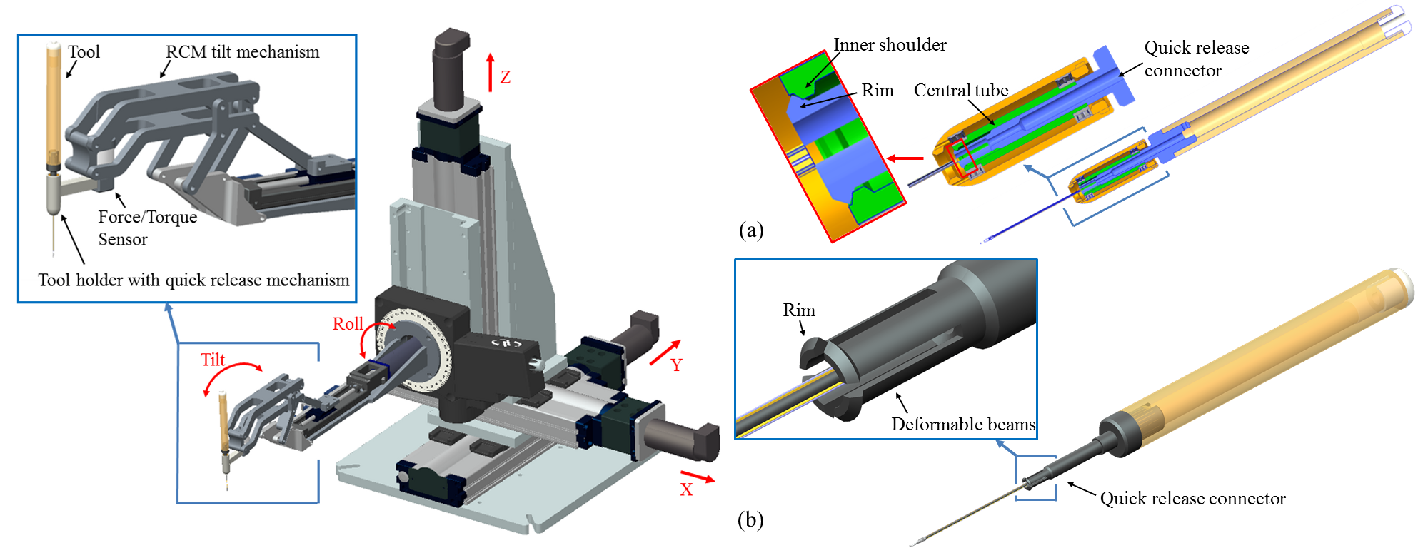
The previously developed SteadyHand Eye Robot has been extensively used in in vivo experiments. Several safety and ergonomic limitations observed in the in vivo environment serve as motivation for a novel robot wrist design. The new robot wrist consists of a symmetric remote center of motion (RCM) tilt mechanism and a slim tool holder with a quick release mechanism for the surgical instruments.

The new RCM tilt mechanism offers a symmetric structure that can be used by both left- and right-handed users, and also enables a two-robot configuration for bilateral manipulation. It can provide a tilt motion range of +/- 45 deg which guarantees sufficient workspace for vitreoretinal surgery. We also analyzed the kinematics of the new RCM tilt mechanism. It demonstrates a fairly linear mapping between the translation of the linear stage and the RCM tilt angle. This can facilitate implementation of more sophisticated control algorithms for micromanipulation. The stiffness of the new RCM tilt mechanism is evaluated with both FEA simulation and experiments. The simulation and the experiment results are consistent with a constant offset. The actual stiffness measured in the experiment is 48.54 N/mm at the negative extreme position, 20.92 N/mm at the center position and 51.02 N/mm at the positive extreme position.
The tool holder enables several important features in a very compact package. It provides axial fixation of the tool while allowing the DOF of rotation about tool axis (spin). It can be easily attached and detached from the robot wrist using a set screw. This enables the separation between sterilizable and non-sterilizable parts that is essential for clinical use of the Eye Robot. The quick release mechanism allows the emergency retraction of the tool from the tool holder, as well as a convenient way to switch between different surgical tools. We designed one “soft” tool with low release force threshold of 2-3 N and one “hard” tool with high release force threshold of 5-6 N.
Robot Assisted Vein CannulationFreehand Vein Cannulation
Robot Peeling Robot Peeling
Tele-Operation with the DaVinci Master Console
Recently we have built a quick tool release mechanism as well as updated the mechanical RCM mechanism. New RCM is more compact and symmetrical. We have been incorporating smart instruments into the robotic system. The force measurements obtained using our micro-sensing instruments are used to perform basic tissue characterization and augment existing control algorithms. We developed a novel control method we call augmented cooperative control, where the operator can alter the direction of a global limit, while the robot guides the tool towards a locally minimum resistance direction. Preliminary experiments with raw eggs both show the capabilities of our platform in characterizing the delicate inner shell membrane, and test our new control algorithm in membrane peeling operations.
The next series of experiments will focus on membrane peeling experiments on real eyes. Quantifying range of forces would enable us to 1) define the range of forces we are dealing with, and develop warning signals for the surge-on if the forces are exceeded, 2) compare human free-hand and robotic assisted performance for the intended application, 3) design virtual models to be used as the basis of a surgical training set.
We are conducting a human subject comparison study of freehand and robot-assisted retinal vein cannulation using the chick chorioallantoic membrane as an eye phantom. We hope to show that using the robot can increase success rate of cannulation and increase the time the micropipette is maintained in the retinal vein during infusion. We also hope to quantify performance differences between populations by recruiting both surgeons and non-surgeons for our comparison study.
We will do more extensive ergonomics research in a surgical suite designed for eye surgery.
"Junking the Joystick" - ASME Mechanical Engineering Magazine Article on how “Medical researchers have discovered that the best way to operate microscale devices is through intuitive controls.” 2007
"Snake-Like Robot and Steady-Hand System Could Assist Surgeons" - Johns Hopkins University News Release 2006. Associated videos
"Hopkins Reports Snake-Like Robot, Steady-Hand System" MedGadget Article 2006
NSF: Robots help surgeons transcend human limits. 2010
SPECTRUM: Special Report: ROBOTS FOR REAL : Surgeons and Robots Scrub Up, 2009