Contact Us
CiiS Lab
Johns Hopkins University
112 Hackerman Hall
3400 N. Charles Street
Baltimore, MD 21218
Directions
Lab Director
Russell Taylor
127 Hackerman Hall
rht@jhu.edu
Problem
Prostate cancer is the most common male cancer in North America. MRI-guided prostate interventions (biopsy and brachytherapay) is proved to provide better results due to the superior image quality of MRI. Up to date, a handful of robotic systems have been introduced to assist manual MRI-guided percutaneous prostate needle placement. These systems range from fully manual systems to fully actuated systems. However, no system has been able to meet all clinical requirements mainly due to insufficient system integration with MRI and exclusion of clinicians from the procedure by taking autonomous approach in robot design. Hence, a clinically approved MRI-compatible assistant robot which can perform prostate intervention more accurately and reliably with less trauma to patient is highly in demand.
Solution
So far, we have developed two versions of pneumatic robot for MRI-guided needle placement. Figure 1 shows the firs robot prototype with two scissor mechanisms in horizontal and vertical plane. Unlike the standard manual procedure in which a grid of template is used to guide surgical needles, the robot can also provide yaw and pitch angulation in order to cover the entire prostate capsule and also to avoid pubic arc/urethra interference.
Figure 1. The first robot manipulator consisting of two scissor mechanisms while doing phantom experiment (left side). Vertical scissor mechanism (right side)
Due to the high actuation force required to move the robot which can negatively affect robot accuracy as well, a parallel kinematic architecture was designed later as shown in Figure 2. The pyramid shape of the robot will optimally use the space under the patient's legs.


Figure 2. The second robot manipulator- a 4-DOF parallel manipulator- while doing phantom experiment (left side). Robot closeup view (right side)
System Components
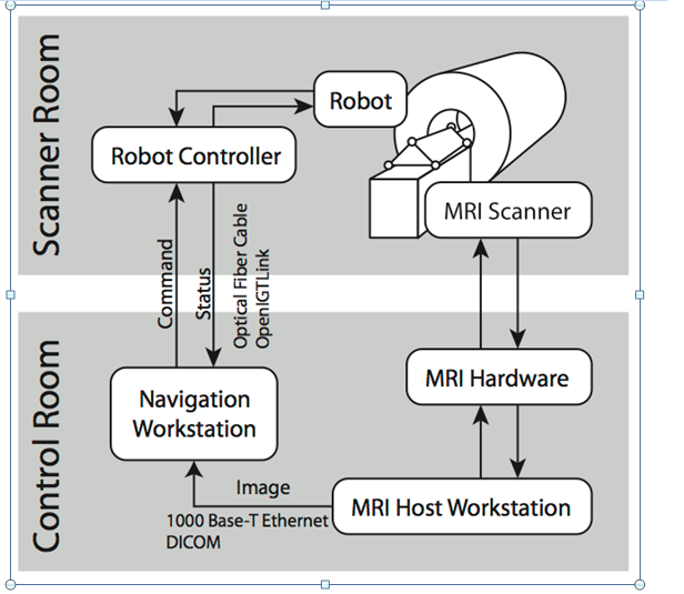
 Figure 3. Diagram of system components and information flow. Red sharp rectangles are explained in future plan
In MR suite (Figure 3), the patient, the robot and the robot controller are placed. The pneumatic robot needs air to run; hence, the controller is connected to the medical air supply which is available in hospital. The controller sends the commands to the robot and gets position feedback from the robot optical encoders. The DC power is transmitted through the patch panel which is an Electro Magnetic (EM) filtered panel specifically designed for this reason in MR room. In the control suite(right hand side), there is scanner console to control the MRI machine. There is another workstation on which 3D Slicer (http://www.slicer.org/) is installed and named as “planning workstation”. 3D Slicer is an interface between the scanner console and robot controller as robot controller cannot directly communicate with scanner console. In 3D Slicer, prostate image is downloaded from scanner console. Target positions are selected by clinician in this software and sent as commands to the robot controller. A local network should therefore be established among the planning workstation, scanner console and the robot controller.
Results
The robot performance is evaluated in terms of MRI compatibility, targeting accuracy and clinical workflow feasibility.
Third generation of the robot
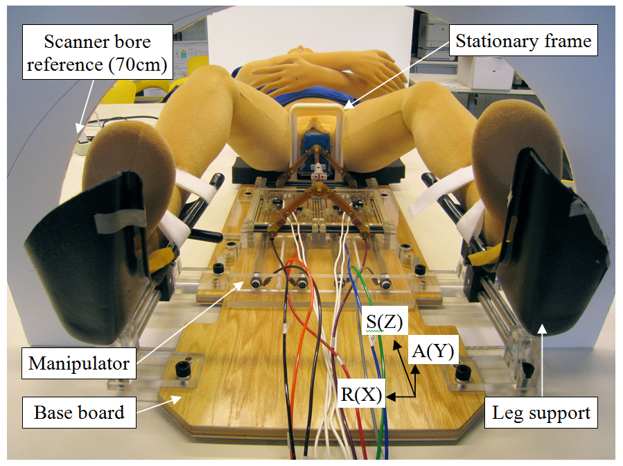
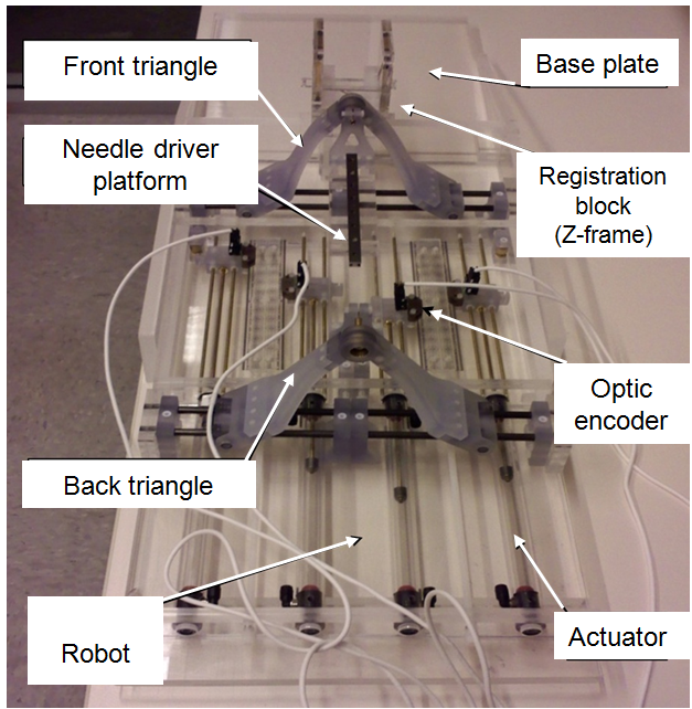
In the third prototype of such robotic system, we have designed and developed a minimally invasive surgical manipulator for MRI-guided transperineal prostate interventions. The proposed manipulator takes advantage of 4 sliders actuated by the MRI compatible piezomotors and rotary encoders. Severe constraints for the MRI-compatibility to hold the minimum artifact on the image quality and dimensions restraint of the bore scanner shadow the design procedure. Regarding the constructive design, the manipulator kinematics has been optimized and the effective analytical needle workspace is developed and followed by proposing the workflow for the manual needle insertion. The introduced robotic manipulator herein is aimed for the clinically prostate biopsy and brachytherapy applications. Here are the summary of the anticipated robot specifications:
• 4 actuated DOF (X, Y, Rx, Ry) • Range of motion: X= ± 35 mm; Y = 130±35 mm, Rx, Ry = ±10° • Resolution of the robot’s motion 0.1 mm or better • Robot can travel through its range of motion within 30 seconds • Accuracy at the needle’s tip in air ±0.5 mm or better, in X-Y plane • Needle insertion’s accuracy at the needle’s tip in air ± 1.5 mm or better • Sufficient stiffness and no backlash • Modular design for manual and tele-operated needle insertion • Compliance with clinical workflow and safety regulations
Results
The robot’s accuracy, repeatability, and MRI compatibility have been evaluated resulting in sustainability and precision of the robot: